| Table of Contents | |
|
Case Report
| ||||||
| Creatine phosphokinase did not increase after on-pump beating heart coronary artery bypass graft early after acute myocardial infarction | ||||||
| Tamaki Takano1, Takamitsu Terasaki1, Yoshinori Ohtsu2, Yuko Wada2, Tatsuichiro Seto2, Daisuke Fukui2 | ||||||
|
1MD, Nagano Red Cross Hospital, Department of Cardiovascular Surgery, Wakasato, Nagano, Japan.
2MD, Shinshu University School of Medicine, Department of Cardiovascular Surgery, Asahi, Matsumoto, Japan. | ||||||
| ||||||
|
[HTML Abstract]
[PDF Full Text]
[Print This Article]
[Similar article in Pumed] [Similar article in Google Scholar] |
| How to cite this article |
| Takano T, Terasaki T, Ohtsu Y, Wada Y, Seto T, Fukui D. Creatine phosphokinase did not increase after on-pump beating heart coronary artery bypass graft early after acute myocardial infarction. Edorium J Cardiothorac Vasc Surg 2015;2:30–35. |
|
Abstract
|
|
Aims:
Emergency coronary artery bypass grafting (CABG) is sometimes mandatory although operative mortality is high within 24 hours after the onset of acute myocardial infarction (AMI). We have used on-pump beating heart (OPBH) CABG to reduce injury to non-infarcted myocardium during CABG. We compared clinical outcomes of OPBH with those of conventional CABG with cardiac arrest (OPCA), both performed within 24 hours after the onset of AMI.
Methods: Twenty-two patients were enrolled in this study. Patients' basic characteristics, operative procedure, in-hospital mortality, morbidity and changes in creatine phosphokinase (CPK) and myocardial subset of CPK (creatine phosphokinase-myoglobin binding, CPK-MB) and ejection fraction (EF) were retrospectively obtained by reviewing hospital records. Results: In the OPCA group, postoperative increases were seen in mean CPK (p=0.03) and in CPK-MB (p=0.03). In the OPBH group, postoperative decreases were seen in mean CPK (p=0.43) and in CPK-MB (p=0.07). Ejection fraction increased postoperatively in both groups, although the increase in OPCA was not significant. No statistically significant differences were found in mortality and morbidity between OPCA and OPBH. Conclusion: In our study, OPBH did not increase CPK and CPK-MB in early AMI treatment, suggesting that OPBH may reduce myocardial injury during CABG performed very early after AMI although further studies including randomized trial are warranted. | |
|
Keywords:
Acute myocardial infarction, Beating heart, Coronary artery bypass graft, Creatine phosphokinase
| |
|
Introduction
| ||||||
|
Operative mortality is high (7.9–15.5%) when coronary artery bypass grafting (CABG) is performed within 24 hours after the onset of acute myocardial infarction (AMI) [1] [2] . However, emergency CABG is required early after AMI when the culprit lesion is located in an unprotected left main trunk or a position that is anatomically unfavorable for percutaneous coronary intervention (PCI), although the advantages of PCI for the treatment of AMI are widely recognized. Previous authors have suggested that on-pump beating heart (OPBH) CABG might be useful in AMI treatment because it did not cause ischemia to non-infarcted myocardium. Therefore potentially minimizing inadvertent injury or edema or both in the setting of AMI [3] [4] [5] [6]. However, it is difficult to determine whether OPBH or conventional CABG with cardiac arrest (OPCA) is favorable early after AMI because patient characteristics, hemodynamic status, and timing of the operation differ in each AMI setting. We selected patients who were within 24 hours after the onset of AMI and compared clinical outcomes of OPBH with those of OPCA. | ||||||
|
Materials and Methods
| ||||||
|
We performed 31 solitary CABG procedures for AMI from March of 2007 to April of 2013 at our institution. Of 31 cases, skin incision was started within 24 hours after the onset of AMI symptoms in 22 patients. OPBH was performed in 10 cases and OPCA in 12 cases. Diagnosis of AMI was made by ST-segment elevation on electrocardiogram (ECG); increases in troponin-T, creatine phosphokinase (CPK), and myocardial subset of CPK (creatine phosphokinase-myoglobin binding, CPK-MB); and reduced kinesis of the left ventricle on ultrasonic echocardiography. Patients' basic characteristics; operative procedures; in-hospital mortality, morbidity, duration of IABP support, intubation period, ICU and hospital stay after the surgery as well as changes in CPK, CPK-MB and echocardiographically determined ejection fraction (EF) were retrospectively obtained by reviewing the hospital records of 22 patients. Cardiopulmonary bypass (CPB) was established via cannulation of the ascending aorta and right atrium after a standard median sternotomy. OPCA was performed from 2007 to 2009 whereas OPBH was done after 2010. The four surgeons participating in this study were on-call and were randomly called to perform emergency CABG. Cardiac arrest in OPCA was achieved using intermittent antegrade crystalloid cardioplegia (Miotector; Terumo, Tokyo, Japan). The only crystalloid cardioplegia was delivered through aortic root catheter every 30–60 min, and blood cardioplegia and terminal cardioplegia were not used. Rectal temperature was maintained 34–36°C throughout CPB, and adequate doses of dopamine and dobutamine were continuously administered beginning immediately upon CPB weaning. Intra-aortic balloon-pump (IABP) support continued until the patient became hemodynamically stable and CPK peaked in patients who were started on IABP preoperatively. We took blood samples 3–6 hours before surgery and 1 hour after its completion. Ejection fraction was measured with ultrasonic cardiography before surgery and 10 -11days after IABP and catecholamine were weaned off. Patients' basic characteristics were compared between OPCA and OPBH groups using t-test or chi-square test, whereas CPK and CPK-MB was compared between groups and between preoperative and postoperative in each group by Wilcoxon rank test or Mann-Whitney U-test using PASW Statistics for Windows, Version 18 (SPSS Inc., Chicago, IL, USA). Statistical significance was assumed to be p<0.05. | ||||||
|
Results | ||||||
|
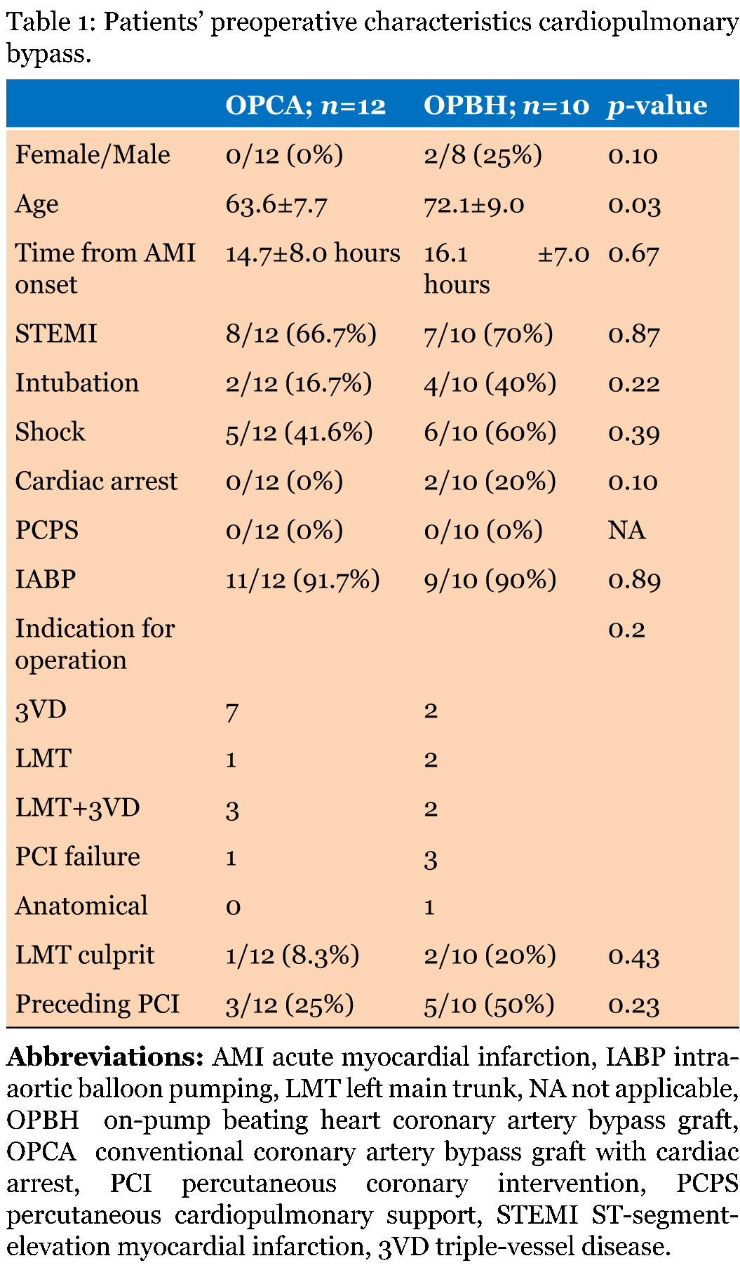
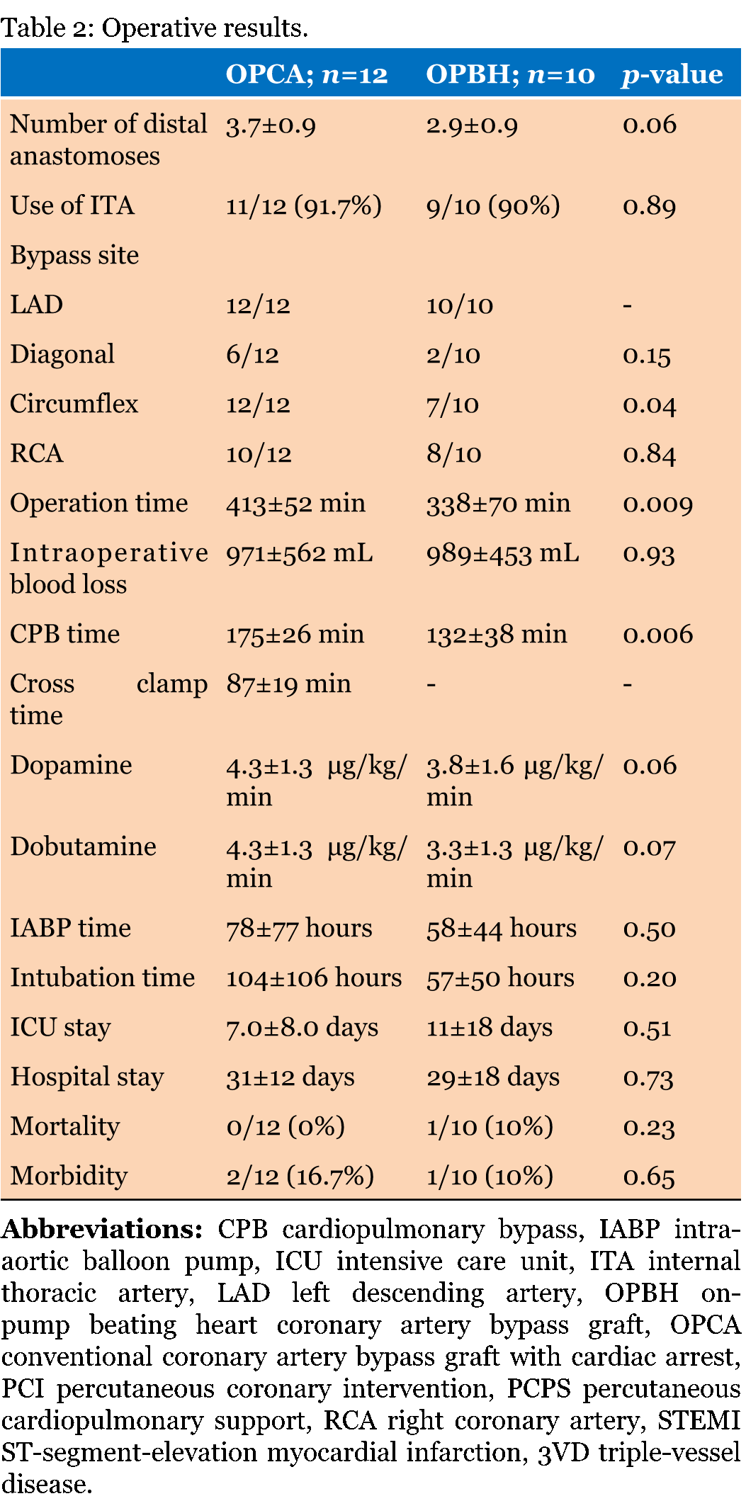
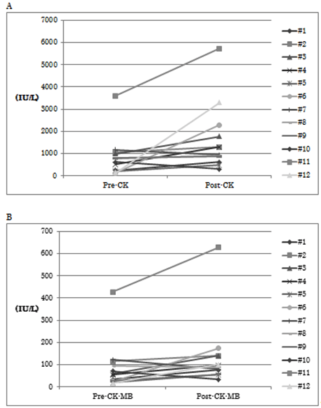
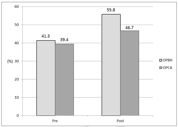
Patient characteristics are given in Table 1. The OPBH patients were older than the OPCA patients. Time interval between the onset of AMI symptoms and skin incision was 14.7±8.0 and 16.1 ±7.0 hours in the OPCA and OPBH groups, respectively. Two patients in the OPBH group presented in cardiac arrest and received cardiopulmonary resuscitation before arrival at the hospital. Table 2 describes operative results. CPB and operative durations were shorter in the OPBH group than in the OPCA group, and there were fewer distal anastomoses and smaller doses of dopamine and dobutamine required at CPB weaning in the OPBH group than in the OPCA group, although these differences did not reach statistical significance. No mortality was observed in the OPCA group, but one patient (9.1%) in the OPBH group died of sepsis 64 days after surgery. In the OPCA group, neurological deficit was observed in one patient, and one patient suffered from low cardiac output syndrome during the surgery and required percutaneous cardiopulmonary support. Continuous hemofiltration was temporarily used in two patients in the OPCA group. However, no statistically significant differences were found in these morbidities between groups. The doses of dopamine and dobutamine required at CPB weaning were not significantly different between the OPCA and OPBH groups. CPK and CPK-MB increased after surgery in 9 of 12 OPCA patients but decreased after surgery in 8 of 10 OPBH patients compared with before surgery (Figure 1) and (Figure 2). In the OPCA group, significant increases were seen in mean CPK (852±936 versus 1,647±1,532 IU/L, p=0.03) and CPK-MB (88±113 versus 139±159 IU/L; p=0.03). In the OPBH group, mean CPK decreased after the surgery (1,562±1,523 versus 1,467±1,254 IU/L, p=0.43), as did CPK-MB (191±196 versus 130±111 IU/L; p=0.07), although the differences were not statistically significant. EF increased postoperatively in both groups, but the increase achieved statistical significance only in the OPBH group (Figure 3). | ||||||
| ||||||
| ||||||
| ||||||
| ||||||
|
| ||||||
|
| ||||||
|
Discussion
| ||||||
|
Percutaneous coronary intervention is the preferred treatment in AMI, but emergency CABG is performed when the culprit lesion is in an unprotected left main trunk or in an inaccessible position, or there are mechanical complications associated with AMI. Clinical outcomes of PCI were shown to be favorable in our previous study [7]. A recent review has demonstrated that PCI has a lower incidence of periprocedural and short-term morbidities but higher rates of repeat revascularization after PCI. CABG has a better durability with a lower rate of mid-term repeat revascularization [2] [8]. In a recent study, off-pump CABG applied to acute coronary syndrome without ST-segment elevation was demonstrated to have lower incidences of bleeding and myocardial infarction but higher rates of re-intervention than OPCA [9]. Off-pump CABG does not unload the ventricles, and cardiac output during surgery depends on cardiac function of the diseased heart. When we performed CABG in patients with AMI, OPBH did not increase, but apparently decreased, CPK and CPK-MB, whereas both CPK and CPK-MB increased after OPCA for AMI. A previous paper reported that myocardial lymph flow was maintained with minimal contraction during CPB, although it decreased to less than 30% of baseline during cardioplegic arrest during CPB, and myocardial edema measured after CPB with minimal contraction was significantly less than that with cardioplegic arrest [10]. Cardioplegic arrest by itself also injured non-infarcted myocardium and resulted in lower oxygen consumption, lower adenosine triphosphate and higher lactate concentrations, and higher water content compared with before arrest 4 days after AMI in an animal model [11]. Alwan et al. reported that troponin-I and CPK-MB release was lower in OPBH than in OPCA after elective CABG [12]. Our results were consistent with their results, although our study included only emergency cases, and myocardial contraction without cardioplegic arrest resulted in less CPK and CPK-MB release in our study. It is well known that changes in CPK and CPK-MB are time dependent after AMI. This study included only patients who underwent skin incision for CABG within 24 hours after onset of AMI symptoms. A previous paper reported that release of CPK-MB was smaller in OPBH than in OPCA in unstable angina patients, whereas the release of CPK-MB was not different between OPBH and OPCA in AMI patients, and concluded that the change in CPK-MB was mainly attributable to the timing of surgery after AMI onset [1]. However, another paper demonstrated OPBH to result in significantly lower CPK-MB compared with OPCA when CABG was performed 9.4 to 10.4 hours after AMI onset [3]. Our study supports their results and suggests that OPBH may reduce myocardial injury during CABG very early after AMI. Operative time and CPB time were shorter in OPBH than in OPCA. The number of distal anastomoses was smaller in OPBH than in OPCA (2.9±0.9 versus 3.7±0.9, respectively), and the shorter operative and CPB durations were attributed to this fewer anastomosis. The doses of dopamine and dobutamine required at CPB weaning were smaller in OPBH than in OPCA, although the differences were not statistically significant. However, OPBH may induce less myocardial injury and improve clinical outcomes of CABG after AMI. Reported mortality of OPBH after AMI varies widely, from 2.6 to 19.3% [2] [3] [6], because the number of patients was small. Our study also included only 10 cases of OPBH, and further study is warranted to clarify the mortality of OPBH with a larger sample size. Three patients suffered from AMI due to a left main trunk lesion, two of whom presented with shock and were intubated before CABG. Neither patient could be weaned from CPB without IABP and percutaneous cardiopulmonary support. One patient who underwent OPCA had a large cerebral infarction and required transfer to a rehabilitation hospital after 27 days of ICU stay and 68 days of hospitalization following CABG. Another patient underwent OPBH and died of sepsis after remaining on a mechanical ventilator for 64 days in the ICU although PCPS and IABP were weaned off. CABG for AMI patients with shock due to a left main trunk lesion is still considered challenging. | ||||||
|
Conclusion
| ||||||
|
On-pump beating heart did not increase creatine phosphokinase (CPK) early after acute myocardial infraction (AMI) and required less catecholamine support during cardiopulmonary bypass (CPB) weaning than did coronary artery bypass grafting (CABG) with cardiac arrest (OPCA). These results suggest that OPBH may reduce myocardial injury compared to conventional OPCA early after AMI. We need more study from larger population and randomized study to clarify the clinical benefit of OPBH. | ||||||
|
References
| ||||||
| ||||||
|
[HTML Abstract]
[PDF Full Text]
|
|
Author Contributions:
Tamaki Takano – Substantial contributions to conception and design, Acquisition of data, Analysis and interpretation of data, Drafting the article, Revising it critically for important intellectual content, Final approval of the version to be published Takamitsu Terasaki – Analysis and interpretation of data, Revising it critically for important intellectual content, Final approval of the version to be published Yoshinori Ohtsu – Analysis and interpretation of data, Revising it critically for important intellectual content, Final approval of the version to be published Yuko Wada – Analysis and interpretation of data, Revising it critically for important intellectual content, Final approval of the version to be published Tatsuichiro Seto – Analysis and interpretation of data, Revising it critically for important intellectual content, Final approval of the version to be published Daisuke Fukui – Analysis and interpretation of data, Revising it critically for important intellectual content, Final approval of the version to be published |
|
Guarantor of submission
The corresponding author is the guarantor of submission. |
|
Source of support
None |
|
Conflict of interest
Authors declare no conflict of interest. |
|
Copyright
© 2015 Tamaki Takano et al. This article is distributed under the terms of Creative Commons Attribution License which permits unrestricted use, distribution and reproduction in any medium provided the original author(s) and original publisher are properly credited. Please see the copyright policy on the journal website for more information. |
|
|