| Table of Contents | |
|
Case Report
| ||||||
| Unusual case of isolated tracheal tear due to blunt neck trauma: Considerations of management | ||||||
| Derek Lim1, Jaime Yun2, Mark Genovesi2, Syed Ali Rizvi3 | ||||||
|
1MS-4, New York Institute of Technology College of Osteopathic Medicine.
2MD, NYU Lutheran Medical Center Department of Surgery. 3DO, NYU Lutheran Medical Center Department of Surger. | ||||||
| ||||||
|
[HTML Abstract]
[PDF Full Text]
[Print This Article]
[Similar article in Pumed] [Similar article in Google Scholar] |
| How to cite this article |
| Lim D, Yun J, Genovesi M, Rizvi SA. Unusual case of isolated tracheal tear due to blunt neck trauma: Considerations of management. Edorium J Cardiothorac Vasc Surg 2015;2:36–41. |
|
Abstract
|
|
Introduction:
Tracheobronchial injuries (TBI) are rare occurrences that must be quickly diagnosed and managed appropriately. TBIs are usually caused by erroneous intubation or bronchoscopy. Patients may initially present with dyspnea or dysphonia, but can quickly develop subcutaneous emphysema and a low oxygen saturation. Post-traumatic tracheobronchial tears are difficult to diagnosis and treatment is often delayed due to its insidiousness. Hallmark signs and radiological findings are important features in the management and survival of these patients.
Case Report: A 59-year-old male sustained a blunt injury to the neck by falling forward onto his bicycle handle bar. He presented with dyspnea, massive subcutaneous emphysema, bilateral pneumothoraces and subsequently found to have anterior and posterior tracheal tears measuring 2 cm on bronchoscopy. The patient was initially managed conservatively but on secondary survey he required surgical repair of the lacerations. Conclusion: Surgical and conservative treatments of post-traumatic tracheobronchial injuries were compared over the past thirty-one years through six different studies. This overview has led to the finding that the increase in conservative treatment have led to a decrease in mortality and morbidity when compared to surgical treatments. | |
|
Keywords:
Blunt neck trauma, Tracheobronchial injury, Subcutaneous emphysema
| |
|
Introduction
| ||||||
|
Tracheobronchial injuries commonly occur due to iatrogenic reasons. Approximately, 1:20, 000 intubation difficulties such as poor insertion or hyperinflation of the ETT cuff result in a longitudinal tear at the points of weakness within the cartilaginous rings of the trachea [1][2]. These tracheal tears are usually located in the posterior wall [1]. Other causes may be due to neck or chest trauma, inhalation of smoke or aspiration of liquids [2]. Although TBIs are rare, the patient must be promptly diagnosed and aggressively managed or else the patient may quickly decompensate [1][2][3][4][5]. Fiberoptic bronchoscopy is the gold standard in diagnosing these injuries [1] [4] [5][6]. Post-traumatic tracheobronchial injuries are less common but should be treated similarly. Studies have shown that 76–80% of post-traumatic TBIs occur on the anterior aspect of the trachea and approximately 2–2.5 cm above the carina [3]. Blunt traumas, defined as high velocity, high impact forces, account for a majority of these cases, of which motor vehicle accidents are the main culprit [2] [4]. Classic signs and symptoms of a TBI include subcutaneous emphysema, stridor, dyspnea and dysphonia [5][6]. Treatment can vary depending on the severity of the injury. Studies have shown positive results in both surgical and conservative treatments [1] [2] [3] [4] [5] [6]. This report presents a case of an anterior tracheal tear with extensive subcutaneous emphysema due to blunt neck trauma. The focus of this case report is to demonstrate the management of tracheal tears, including prompt intubation, visual examination via fiberoptic bronchoscopy and proper treatment plan assessment. | ||||||
|
Case Report
| ||||||
|
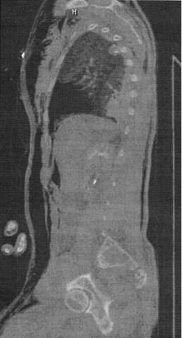
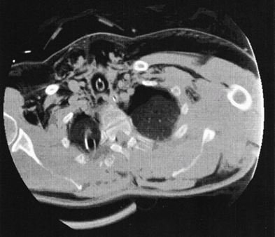
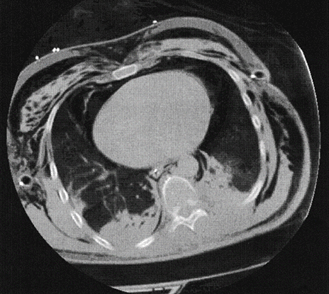
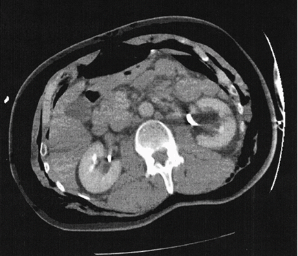
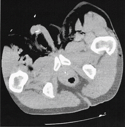
A 59-year-old male with no significant past medical history, arrived in the trauma bay boarded and collared by EMS with supplemental oxygen administered via a non-rebreather mask. EMS reported that he was biking along the road when he suddenly hit a pothole causing him to jolt forward, striking his neck against the handlebars. The patient presented with a patent airway but complained of extreme difficulty breathing. His initial vitals upon arrival were temperature 97.9°F, blood pressure 236/113 mmHg, pulse 102 beats per min, respirations 22 breaths per min, O2 saturation 93%. The patient had a GCS of 15 upon arrival. Upon physical examination, there was crepitus in the face, neck, chest wall, abdomen, extremities and genitals, suggestive of extensive subcutaneous emphysema. Ecchymosis and abrasions were noted on the anterior neck between the cricoid cartilage and sternal notch (Zone 1). An abrasion was also noted on the left upper extremity but was unremarkable. On auscultation there were bilateral decreased breath sounds. There were no other visible lacerations or deformities. Further physical examination was within normal limits. While in the trauma bay, he became unable to phonate, hypoxemic and combative at which point he was immediately intubated by anesthesiology. A 7.5 mm endotracheal tube was inserted without difficulty and no resistance was met upon placement. Post-intubation, the blood pressure decreased to 226/110 mmHg, pulse decreased to 91 beats per min and respirations decreased to 18 breathes per min. Oxygen saturation increased to 98%. Laboratory values drawn in the trauma bay revealed white blood cells 14.3x10³/ul, hemoglobin 15.7 g/dL, hematocrit 47.5%, platelet 252x103/ul, PT 10.6 sec, PTT 21.9 sec, INR 1.0. Arterial blood gas showed pH 7.29, CO2 51 mmHg, PO2 107 mmHg, bicarbonate 24.5 mEq/L. A portable chest X-ray film showed extensive subcutaneous and mediastinal emphysema, along with bilateral pneumothoraces. Bilateral chest tubes were placed which put out minimal amount of serosanguinous fluid. CT scan of the neck showed diffuse mediastinal and subcutaneous emphysema and confirmed the bilateral pneumothoraces (Figure 2). Lateral and abdominal CT imaging showed extensive subcutaneous emphysema and free air under the rectus sheath (Figure 1) (Figure 2) (Figure 3) (Figure 4). CT imaging of the lower extremities also showed some subcutaneous emphysema along with free air in the scrotum (Figure 5). Further imaging ruled out fractures or cranial hematomas. A bronchoscopy and esophagoscopy were performed in the SICU to rule out tracheal and/or esophageal injury. A two centimeter anterior and posterior wall mid-tracheal tear approximately 5 cm above the carina was noted upon exam. ETT was placed distal to the injury. Esophagoscopy did not show any gross injuries of the esophagus. Patient initially remained intubated and was conservatively treated. On hospital day two, the patient's WBC began to increase despite a minimal decrease in the subcutaneous emphysema. A secondary survey of the tracheal tear with the fiberoptic bronchoscope was performed, and it was agreed upon that the patient would undergo a surgical repair. The patient was treated with an anterior neck incision and exploration. The posterior tracheal injury was repaired using simple interrupted sutures. The anterior tracheal injury was debrided and a tracheostomy tube was placed through it. The cuff was deflated for the entire postoperative course. The patient was decannulated on postoperative day nine and discharged home without further complications. | ||||||
| ||||||
| ||||||
| ||||||
| ||||||
| ||||||
|
Discussion
| ||||||
|
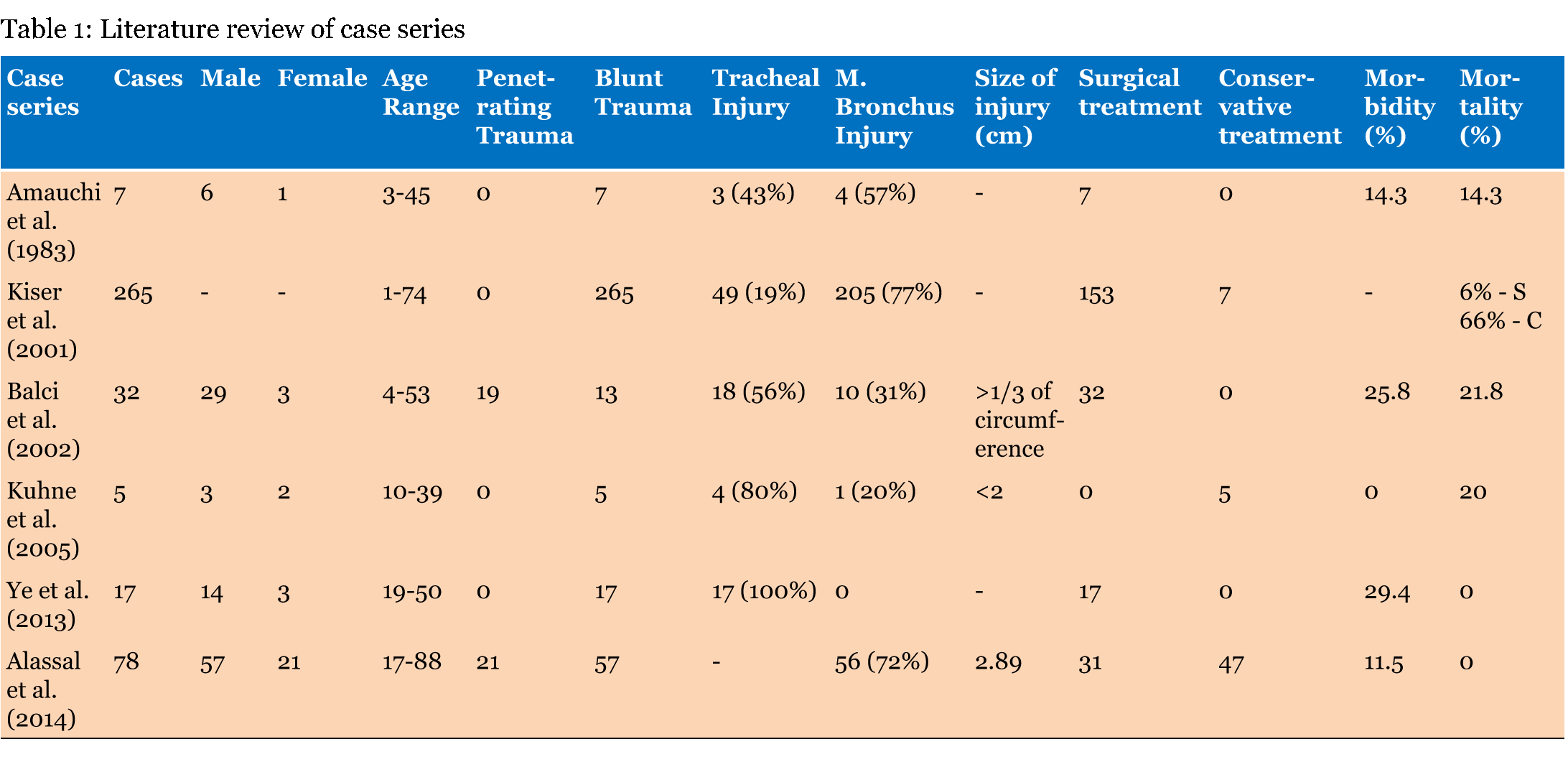
Tracheobronchial injuries have been continuously studied over the past 30 years because of the increase in the number of cases. This can be a result of the increasing number of motor vehicle accidents as well as the advancement in the quality of care during transport by EMS [5]. A recent study by Alassal et al. showed that blunt traumas contributed to 73% of TBIs while a previous study in 2002 by Balci et al. reported blunt traumas contributing to only 41% of TBIs [5] [6]. Management protocols such as securing the airway and diagnostic bronchoscopy are crucial in these patients. Dyspnea, subcutaneous emphysema or difficulty ventilating are clear clinical signs for endotracheal intubation. Pneumomediastinum, pneumothoraces or hemothoraces found on chest X-ray also assist in making the diagnosis [5] [6]. Thoracostomy tubes are also placed to relieve the pneumothorax or hemothorax. However, if the patient's symptoms continue to worsen, a continuous air leak should be suspected and the patient should be taken to the operating room for an emergent flexible bronchoscopy [1] [4] [5][6]. Further management is based upon the findings of the bronchoscopy. Surgical treatment has been shown to be an effective treatment method in repairing TBIs [3] [5][7][8]. In a comprehensive study over a 14-year period by Balci et al., thirty-two patients were reported with either a penetrating or blunt trauma resulting in a complete tracheal transection or tracheal laceration greater than a third of the circumference. The diagnosis was made based upon visual bronchoscopy or obvious clinical signs/symptoms such as subcutaneous emphysema, pneumothorax refractory to chest tube insertion, etc. Their study documented thoracotomy procedures in 59.3% of cases and cervical incisions in 31.2% of cases. Balci et al. concluded that a cervical incision was a superior approach because of adequate visualization of the trachea without the morbidities related to thoracotomies. Non-absorbable sutures were used to repair all the injuries. Some morbidities encountered included atelectasis, pneumonia or tracheal stenosis. The study further reported that when surgical treatment was performed within two hours of trauma, there was a 0% mortality rate compared to a 19.3% mortality rate overall [5]. More recently, conservative treatment has become an effective alternative to surgical treatment [1] [2][4]. In a case study by Kuhne et al., conservative treatment was given to patients who were diagnosed more than three days after admission, in poor physical condition, refused the surgical procedure, did not require mechanical ventilation, minimal subcutaneous emphysema or sustained a tracheal tear less than two centimeters [4]. The patients were intubated with the endotracheal cuff inflated distal to the tear and continually observed via series bronchoscopy to note the healing progress of the tracheal injury. The ETT may be left for five to nine days. Multiple studies have shown the effectiveness of conservative treatment within the past decade. For instance, Gomez et al. discussed the effectiveness in conservative treatment of 17 patients who suffered from a TBI. They concluded that a visual diagnosis within 12 hours contributed to improved outcomes and that conservative treatment should be highly considered in patients suffering from tracheal lacerations less than four centimeters regardless of location or delay in diagnosis [1]. Lampl reported a decrease incidence of tracheal stenosis as well as a mortality rate of less than one percent [2]. Lampl et al., Kuhne et al. and Gomez et al. all reported successful conservative treatment of post-traumatic and iatrogenic tracheal tears. All these studies recommend early visual/clinical diagnosis of the tracheal tear using a flexible bronchoscopy in order to improve the outcome of conservative treatment [1][2] [4][6]. In a literature review of the case series on post-traumatic tracheobronchial injuries and treatments, the morbidity and mortality rates between surgical and conservative treatments were studied. Key factors that may influence the treatment outcome include gender, age, types of trauma, location of trauma and size of injury. Due to these variables, a set guideline for the treatment of TBIs has been difficult to formulate [5][6]. Previously, surgical treatment seemed to be the predominant guideline, but outcomes included increased morbidities such as bronchial stenosis, atelectasis and hoarseness. More recently, conservative treatment has been considered an alternative to surgical treatment. It has led to a decrease in mortality from 2001, in which Kiser et al. reported a mortality rate of 66% to Kuhne et al., in 2005, who reported a mortality rate of 20%, and to Alassal et al., in 2014, who reported a mortality rate of 0%. In addition, Alassal et al. reported a morbidity rate of 11.5% after conservative treatments, which is a significant decrease from Ye et al. who reported a morbidity rate of 29.4% post-surgical treatment [3] [4] [5] [6] [7] [8] (Table 1). | ||||||
| ||||||
|
Conclusion
| ||||||
|
This case report demonstrated a rare occurrence of post-traumatic tracheobronchial tear, its management and its treatment. Post-traumatic tracheobronchial injuries are very often delayed in diagnosing because of its subtle and indolent process, taking about seven days and because of the distraction of other more obvious injuries. Noticing clinical and radiological signs are important in the management of these patients. Prompt intubation and proper visualization via bronchoscopy, assist in determining whether to treat the patient conservatively or surgically. Despite the extent of subcutaneous emphysema upon physical examination, this patient was noted to have two 2 cm anterior and posterior tracheal tears. Many studies in the past decade proved the effectiveness of conservative treatment for tracheobronchial injuries and while the rate of morbidity and mortality are increased in surgical repair, it may be a necessary procedure based on the extent of the injury. | ||||||
|
References
| ||||||
| ||||||
|
[HTML Abstract]
[PDF Full Text]
|
|
Author Contributions
Derek Lim – Substantial contributions to conception and design, Acquisition of data, Analysis and interpretation of data, Drafting the article, Revising it critically for important intellectual content, Final approval of the version to be published Jaime Yun – Analysis and interpretation of data, Revising it critically for important intellectual content, Final approval of the version to be published Mark Genovesi – Analysis and interpretation of data, Revising it critically for important intellectual content, Final approval of the version to be published Syed Ali Rizvi – Analysis and interpretation of data, Revising it critically for important intellectual content, Final approval of the version to be published |
|
Guarantor of submission
The corresponding author is the guarantor of submission. |
|
Source of support
None |
|
Conflict of interest
Authors declare no conflict of interest. |
|
Copyright
© 2015 Derek Lim et al. This article is distributed under the terms of Creative Commons Attribution License which permits unrestricted use, distribution and reproduction in any medium provided the original author(s) and original publisher are properly credited. Please see the copyright policy on the journal website for more information. |
|
|