 |
Case Report
Pediatric Cardiac hydatid cysts: A fatal manifestation of the disease
1 Erbil Cardiac Center, Kurdistan, Iraq
2 Sulaimani Teaching Hospital, Sulaimani, Kurdistan, Iraq
3 University of Sulaimani, Old Campus, Sulaimani, Kurdistan, Iraq
4 Kscien Organization for Scientific Research, Azadi Bulding, Sulaimani, Kurdistan, Iraq
Address correspondence to:
Fahmi H. Kakamad
Sulaimani Teaching Hospital, Sulaimani,
Iraq
Message to Corresponding Author
Article ID: 100016C04MM2018
Access full text article on other devices

Access PDF of article on other devices


How to cite this article
Mahmood M, Yaldo FF, Kakamad FH, Abdulla K, Jaff A. Pediatric Cardiac hydatid cysts: A fatal manifestation of the disease. Edorium J Cardiothorac Vasc Surg 2018;5:100016C04MM2018.ABSTRACT
Introduction: Hydatid cysts affecting myocardium is an extremely rare condition. The aim of this study is to report a rare case of cardiac hydatid disease presented with heart failure and a brief review of the literature.
Case Report: A 14-year-old girl, living in an endemic area, presented with severe pulmonary hypertension and right side heart failure secondary to multiple cardiac and pulmonary hydatid cysts diagnosed by echocardiography and chest computed tomography. Intraoperatively, there was mild pericardial effusion, the cysts diffusely affected myocardium in different areas. A process of femoral arterial cannulation, with bicaval cannulation and cardioplegic arrest, was commenced. The contents of the cysts were aspirated and the cavities were washed with povidone iodine. Following cross-clamp removal, the patient could not be weaned off bypass and death declared on the table.
Conclusion: Cardiac hydatid cyst is a very rare variant of the disease; it may present in various scenarios. Surgical intervention is the only hope of survival.
Keywords: Death, Echinococcus granulosus, Myocardium, Parasite
INTRODUCTION
Hydatis, in Greek, means a drop of water, an expression that refers to the fluid containing cysts brought about by the larval stage of Echinococcus granulosus, in which the human acts as an accidental intermediate host [1],[2]. Upon arrival of the embryo to the liver, it either settles there or bypasses it through the lymphatics or systemic circulation to reach the lungs and other organs. The most commonly affected organs are the liver and lungs 70% and 20% respectively. The other locations include the spleen and kidneys, which account for 2% each [3],[4]. Very rarely, hydatid cyst (HC) affects other organs including chest and abdominal walls, diaphragm, spine, posterior mediastinum and lower limbs [5],[6],[7].
Cardiac involvement is unusual, though the exact percentages are differently recorded in the literature, it is about 0.05-2% and they have been increasingly reported [2], [8],[9]. The embryo makes its path to the myocardium via the coronary circulation. Occasionally, it reaches the right side of the heart through the venous return from there to the left side via patent foramen ovale or through the pulmonary veins.
Despite the discrepancy in reported percentage of different parts of the heart, most of the reports in the literature are in agreement that the most commonly affected part is the left ventricle (LV) followed by the interventricular septum, right ventricle (RV), right atrium (RA) with left atrium (LA) and sinus of Valsalva being the least commonly affected [2],[4],[8],[10].
Due to the absence of uniform clinical presentation, a high index of suspicion is crucial especially in endemic areas like Middle East countries [10]. Early diagnosis with an effective treatment plan can change outcomes dramatically, keeping surgical intervention as a priority even in the asymptomatic cases [2],[4].
In this clinical vignette, a unique case of right-sided congestive heart failure combined with severe pulmonary hypertension secondary to cardiac involvement with hydatid cyst is presented with a brief review of the literature.
CASE REPORT
A 14-year-old girl referred to the cardiac center with severe pulmonary hypertension and right side heart failure (NYHA class III) secondary to multiple cardiac and pulmonary hydatid cysts.
The patient was from a rural area, remote from the city, have had contact with the animal. She recalls the earliest symptoms emerging a year ago in the form of cough, progressive dyspnea inclining into orthopnea and generalized body edema including abdominal distension. Early in the presentation, her symptoms ascribed to the renal cause, as one of her abdominal ultrasound (US) showed prominent pelvic calyceal systems with few internal echoes. Abdominal US revealed severe ascites, pleural effusion, and mild pericardial effusion, echocardiography demonstrated severe right side dilatation, mildly compressing left side with multiple cystic lesions inside the pericardial space posteriorly and apically, one of them was inside the left atrium causing pulmonary venous flow obstruction and pulmonary hypertension with peak gradient of 69 mmHg in one record and 80 mmHg in another, right ventricle systolic pressure was 95 mmHg, with moderate tricuspid regurgitation and normal other valve function.
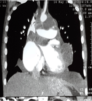
Subsequently, chest computed tomography (CT) scan was performed which showed multiple cysts involving LV myocardium with LA lumen & mediastinum encasing and compressing the pulmonary artery (Figure 1). Upon the above findings, the patient was prepared for urgent operation. Intraoperatively, there were signs of generalized tissue edema, extending to the skin and subcutaneous tissue, confirming the severe right side failure.
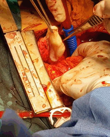
There was mild pericardial effusion, but no pericardial cysts, the cysts were in the wall of the myocardium of the LV, RV extending to outflow tract and aortopulmonary window, LA and pulmonary veins (Figure 2). A process of femoral arterial cannulation, with bicaval cannulation and cardioplegic arrest, was commenced. The contents of the cysts were aspirated, the cavities were washed with povidone-iodine, and cystectomies performed with the closure of the residual cavities.
Lamentably following cross-clamp removal, the patient could not be weaned off bypass and death declared on the table.
DISCUSSION
Cardiac HC, a sparse event yet with dreadful consequences, as described in the literature as a silent bomb [10]. The clinical presentation can be very wide, ranging from asymptomatic to sudden death. Depending on the cardiac structure the cyst invades or compresses and the degree of interference with cardiac function presenting as tamponade, pericarditis, arrhythmias, conduction disturbances, symptoms resembling coronary artery disease, emboli and others [8],[9]. Though the combination of severe pulmonary hypertension and congestive right side failure as it was with the current case has not been reported according to our knowledge. The most probable explanation for this ruinous presentation is attributed to the site of the cysts, as one of them was involving the RV outflow tract causing RV dilatation with failure, the other cysts were involving the LV compressing the pulmonary veins leading to severe pulmonary hypertension. We found one case of cardiac HC involving the pulmonary veins but luckily that patient had a patent foramen ovale which could have act as a pop of that prevented against the development of severe pulmonary hypertension [11].
Another case reported in the literature in which multiple hydatidoses including cardiac cysts led to patient death, in that patient both pulmonary arteries were obstructed due to embolization of the cysts resulting in severe pulmonary hypertension, followed by a rocky postoperative course and eventual death [2].
This might highlight the perils of such presentation in cardiac HC and triggers further investigations in management strategy.
In view of the line of management of cardiac HC, we stand firm with other authors’ opinions that consider the surgical intervention is urgently necessary whenever cardiac hydatid cyst is diagnosed even in asymptomatic cases, as 20% of fatal cases who presented with sudden death were asymptomatic. In consequence, the earliest the action the better the results and the least the undesirable consequences [2],[4],[8],[9].
CONCLUSION
Cardiac HC is a very rare variant of the disease, it may present in various scenarios ranging from asymptomatic one to severe right side failure and pulmonary hypertension. Surgical intervention is the only hope of survival.
REFERENCE
1.
Ohri S, Sachdeva A, Bhatia M, Shrivastava S. Cardiac hydatid cyst in left ventricular free wall. Echo Res Pract 2015 Mar 1;2(1):K17–9. [CrossRef]
[Pubmed]

2.
Ustünsoy H, Akdemir I, Sivrikoz MC, Tahtaci N, Aksoy M, Tunçözgür B. Cardiac hydatid cyst: Report of two cases. Heart Lung Circ 2002;11(2):117–20. [CrossRef]
[Pubmed]
3.
Almutairi A, Al Rajhi S. Case report of hydatid cyst in the pulmonary artery uncommon presentation: CT and MRI findings. Case Rep Radiol 2018 May 15;2018:1301072. [CrossRef]
[Pubmed]
4.
Verma RK, Krishna V, Kumar N, Prakash N, Singh S, Tripathi N. Excision of innumerable hydatid cysts from the myocardium of the left ventricle via the lef thoracotomy along with myoplasty on beating heart. Indian J Thorac Cardiovasc Surg 2018;34(4):496–99. [CrossRef]
5.
Tsigkas G, Chouchoulis K, Apostolakis E, et al. Heart echinococcus cyst as an incidental finding: Early detection might be life-saving. J Cardiothorac Surg 2010 Dec 8;5:124. [CrossRef]
[Pubmed]
6.
Salih AM, Kakamad FH, Hammood ZD, Yasin B, Ahmed DM. Abdominal wall hydatid cyst: A review a literature with a case report. Int J Surg Case Rep 2017;37:154–6. [CrossRef]
[Pubmed]
7.
Salih AM, Kakamad FH, Rauf GM. Isolated hydatid cyst of the diaphragm, a case report. Int J Surg Case Rep 2016;29:130–2. [CrossRef]
[Pubmed]
8.
Akar R, Eryilmaz S, Yazicioglu L, et al. Surgery for cardiac hydatid disease: An Anatolian experience. Anadolu Kardiyol Derg 2003 Sep;3(3):238–44.
[Pubmed]
9.
Yaliniz H, Tokcan A, Salih OK, Ulus T. Surgical treatment of cardiac hydatid disease: A report of 7 cases. Tex Heart Inst J 2006;33(3):333–9.
[Pubmed]
10.
Oraha AY, Faqe DA, Kadoura M, Kakamad FH, Yaldo FF, Aziz SQ. Cardiac hydatid cysts; presentation and management. A case series. Ann Med Surg (Lond) 2018 Apr 7;30:18–21. [CrossRef]
[Pubmed]
11.
Behzadnia N, Hossein-Ahmadi Z, Sharif-Kashani B, et al. Pericardial hydatid cyst in oblique sinus, obstructing all pulmonary veins: A rare presentation. Tanaffos 2013;12(1):78–80.
[Pubmed]
SUPPORTING INFORMATION
Author Contributions
Muhammad Mahmood - Substantial contributions to conception and design, Acquisition of data, Analysis of data, Interpretation of data, Drafting the article, Revising it critically for important intellectual content, Final approval of the version to be published
Fitoon F. Yaldo - Substantial contributions to conception and design, Acquisition of data, Analysis of data, Interpretation of data, Drafting the article, Revising it critically for important intellectual content, Final approval of the version to be published
Fahmi H. Kakamad - Substantial contributions to conception and design, Acquisition of data, Analysis of data, Interpretation of data, Drafting the article, Revising it critically for important intellectual content, Final approval of the version to be published
Kawa Abdulla - Substantial contributions to conception and design, Acquisition of data, Analysis of data, Interpretation of data, Drafting the article, Revising it critically for important intellectual content, Final approval of the version to be published
Avan Jaff - Substantial contributions to conception and design, Acquisition of data, Analysis of data, Interpretation of data, Drafting the article, Revising it critically for important intellectual content, Final approval of the version to be published
Guarantor of SubmissionThe corresponding author is the guarantor of submission.
Source of SupportNone
Consent StatementWritten informed consent was obtained from the patient for publication of this case report.
Data AvailabilityAll relevant data are within the paper and its Supporting Information files.
Conflict of InterestAuthors declare no conflict of interest.
Copyright© 2018 Muhammad Mahmood et al. This article is distributed under the terms of Creative Commons Attribution License which permits unrestricted use, distribution and reproduction in any medium provided the original author(s) and original publisher are properly credited. Please see the copyright policy on the journal website for more information.


